Case Study at The Animal Clinic of St. Petersburg
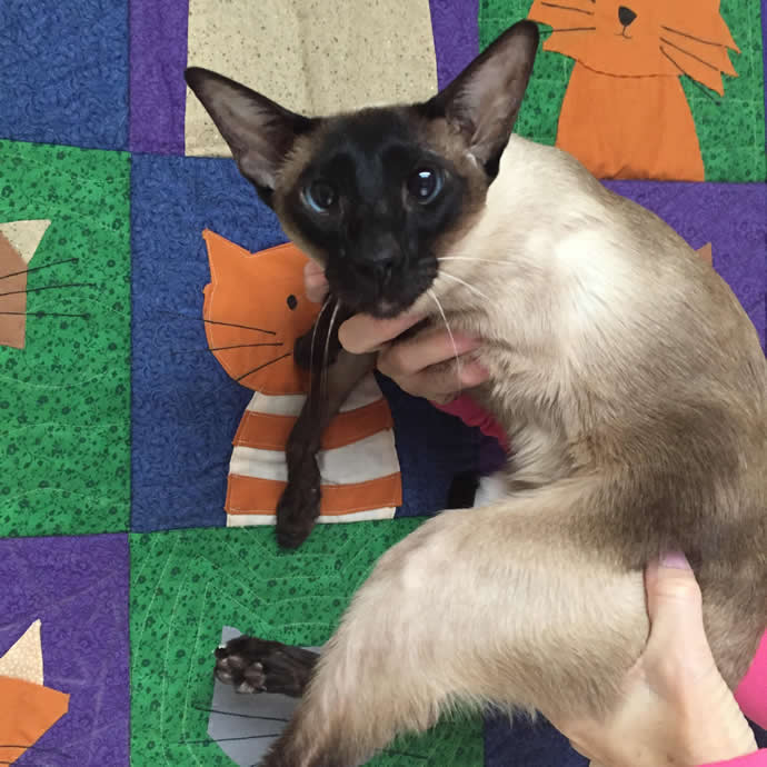
When I was a student (not nearly as many years ago as Dr. Burroughs), we were given example patient stories to study in order to learn various medical conditions and how to manage them. I’m going to give you one very very real life example of an event that occurred recently at The Animal Clinic. This report is what is commonly known in the veterinary profession as a “case study”. This one is about a lovely cat who we will call Simon.

The Animal Clinic St Pete – Case Study: Simon the Cat
This is not his real name and I do have permission from his marvelous owner to share his story with you, but to protect his privacy and not let his fame get too out of hand, we will call him Simon (after “Simon’s Cat”, a ridiculous cartoon that I’ll try to post a link to after you’ve heard all about our Simon).
The Problem
Anyway, Simon has had a long history (way before I came to The Animal Clinic of St Petersburg) of gastrointestinal issues, primarily vomiting. The first time I got to see him, he had a very rough few days of not being able to keep much of anything down and was very thin. Dr. Burroughs decided a barium series was in order because he was concerned about an issue with his pyloric sphincter (now if you were veterinary students you would be sent to the library at this point to research all about the gastrointestinal tract and in particular pyloric emptying. Since you aren’t veterinary students, I’ll just get on with Simon’s story)
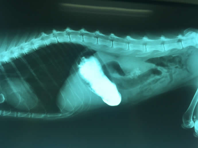
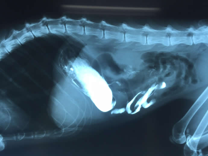
Below are Simon’s radiographic images. The first one with just his stomach filled with the very bright white barium and the lower on with it beginning to move through his intestine.

The Animal Clinic St Pete – Case Study: Simon the Cat – Actual Radiograph Image

The Animal Clinic St Pete – Case Study: Simon the Cat – Actual Radiograph Image
The Diagnosis
What we immediately noticed was that Simon had no problem with his stomach emptying, but did have what looked like a bony obstruction that we assumed was a foreign body blocking his intestinal tract. Now this would actually make sense because our lovely Simon is a bit of an energetic fellow and tends to like to help himself to the trash if he’s not getting what he deems appropriate from his owner. So sure enough, surgery it was for Mr. Simon.
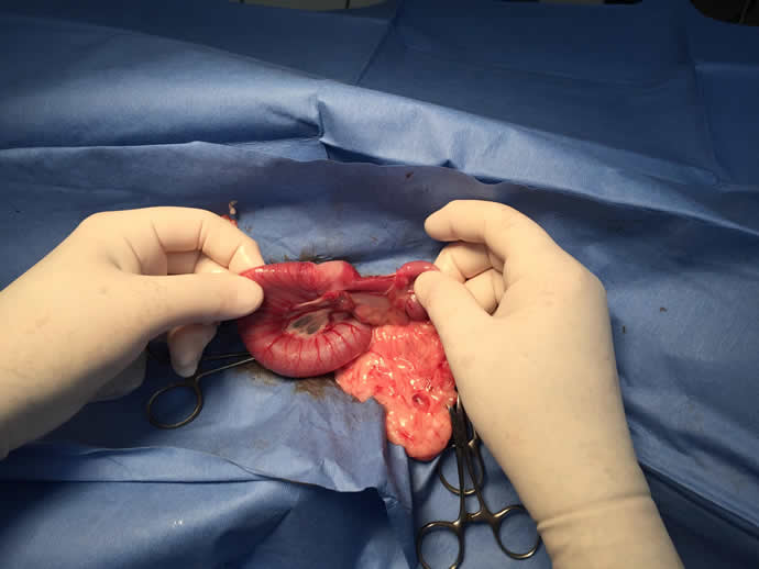
Surgery ended up being quite a surprise because what we thought was a foreign body was actually a section of intestinal tract that had scar tissue so old it had calcified. At some point, poor Simon had had some issue that caused his intestine to form a scar and that tissue had contracted and firmed to the point of being almost as dense as bone, also not letting anything travel much past it. So sure enough we were able to remove that section and reattach the intestine so Simon could pass his food with no issues. I will post surgery pictures at the bottom so any of our more squeamish readers don’t have to see them, but those of you who are contemplating careers in surgery or have strong stomachs feel free to look.
Anyway, after surgery Simon had a bit of a tough time getting back on his feet and required an appetite stimulant as well as anti-inflammatories and antibiotics in order to enable him to realize he could eat without issue. He is now finally feeling great, so great in fact that he recently had to have another radiograph to make sure he didn’t have any chicken bone particles since he had stolen one from the trash.
The moral of Simon’s story is that sometimes even Dr. Burroughs is surprised by what we find in surgery at The Animal Clinic of St Petersburg.
As promised, here’s the Simon’s Cat cartoon weblink: https://simonscat.com/
Warning: These are surgery pictures and may be a little much for some people. The distended intestine is anterior or before the calcified area which is very pale and after or posteriorly the intestine is very small and empty. The drippy looking pale stuff is omentum a lace like fatty tissue in the abdomen it’s all normal.

The Animal Clinic St Pete – Case Study: Simon the Cat – Actual Surgery Image

